
Patellar Luxations
Do you have a toy- or small-breed dog that has intermittent or skipping hindlimb lameness? Then you’ll need to read this!
What is Patellar Luxation?
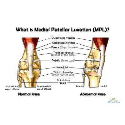
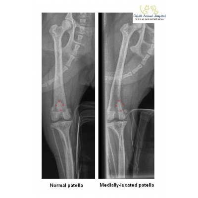
In the normal dog and cat knee, the patella (knee cap) sits in the trochlear groove which is a groove located at the bottom part of the femur/thigh bone. The patella is held in place by tendons and ligaments that keep it relatively stable against the femur when the knee bends (flexes) and extends during movement. Patellar luxation occurs when a pet’s patella slips out of the groove in the femur, causing discomfort, pain, lameness, and knee instability. The patella can slip out towards the inner part of the knee (medial patellar luxation), or towards the outer part of the knee (lateral patellar luxation).
Patellar luxation can occur in one or both knees, and while it is commonly seen in small or toy-breed dogs, many other dog breeds can also be affected. Patellar luxation may also be occasionally observed in cats, although not as commonly as in dogs.
What causes Patellar Luxation?
Patellar luxation may be caused by trauma to the knee; or occur as a developmental abnormality. In the latter situation, patellar luxation is thought to have a genetic basis. During the skeletal development in an immature pet, the trochlear groove fails to develop normally such that the groove is too “shallow” – as a result, with knee movement, the patella slides in and out of the groove. Over time, the sliding motion “wears out” the sides of the trochlear groove, erodes the knee joint’s cartilage and the condition may progress to a stage whereby the patella permanently sits outside of the groove. This may be accompanied by knee arthritis which, develops as a result of abnormal biomechanics (forces of weight-bearing and movement) passing through the unstable knee joint.
What are the Signs of Patellar Luxation?
Clinical signs associated with patellar luxation may not be immediately obvious. Possible clinical signs may include, but are not limited to:
- Knee pain
- Lameness
- “Skipping” gait – whereby pets will occasionally appear to “skip” while walking or running, with the affected leg held up away from the ground. After the knee flexes and extends a few times, some pets will appear to regain the ability to bear weight on the affected limb again. See an example of skipping lameness here.
How is Patellar Luxation Diagnosed?
Patellar luxation is sometimes diagnosed during a routine physical examination when your veterinarian palpates (feels) and manipulates the knee joint. However, if the pet is in a lot of discomfort, your veterinarian may recommend sedation in order to carry out a more thorough examination on your pet’s knee. Radiographs (x-rays) are sometimes also recommended to further evaluate the patella, hindlimb alignment and other structures within the knee, because patellar luxation may sometimes occur in conjunction with other conditions e.g. cranial cruciate ligament tears.
Here at Cahill Animal Hospital, we grade the severity of the patellar luxation on a scale of 1 to 4 as follows:-
- Grade 1 – Intermittent patellar luxation. The patella prefers to sit in the groove of the femur and only slides out of the groove infrequently.
- Grade 2 – Frequent patellar luxation. The patella frequently slides out of the groove but does not immediately go back into the groove. The “skipping” gait is classically seen in pets with Grade 2 luxation.
- Grade 3 – Permanently luxated patella, but able to be manipulated into the groove, but later pops right back out again as soon as the veterinarian releases his/her grip on the patella.
- Grade 4 – Permanently luxated patella, and not able to be manipulated back into the groove by your veterinarian.
The grade of patellar luxation does not necessarily correspond to the pet’s degree of lameness. For example, a dog with a grade 2 luxation may be very lame in comparison to a dog with a grade 3 luxation that appears to walk normally as he might have figured out how to change his gait to compensate for it.
How is Patellar Luxation Treated?
Pets that have been diagnosed with patellar luxation but do not exhibit or show only occasional clinical signs may be conservatively managed with a combination of weight management, exercise moderation, physical therapy, pain relief as needed and/or joint supplements.
Surgery is typically considered for cases where there is a significant degree of lameness. Depending on the patient and severity of the luxation, surgery may involve a combination of deepening the groove where the patella sits in and/or surgical re-alignment of the knee biomechanics. In more severe cases where there is significant hindlimb malalignment as a result of this developmental disorder, a referral to a surgical specialist may be needed to correct the hindlimb deformity. If left untreated, patellar luxation may result in significant damage to the joint, leading to the development of arthritis and other knee conditions such as cranial cruciate ligament rupture.
Remember that every pet is different, and your veterinarian will carefully evaluate your pet and then tailor a treatment plan specifically for your fur friend. You may also read more about patellar luxations here.
The above information is provided for educational purposes only and not intended or implied to be a substitute for professional veterinary medical advice, diagnosis or treatment; and should not be relied on solely as veterinary advice. If you are worried your pet may have patellar luxation, please phone us on (06) 3588675 to book them in for a check over.

